The term Carotids come from the Greek word
karotides. The Carotids are the principal arteries which,
arising from the brachiocephalic arteries, ascend the neck and supply the
head and brain with blood. They exhibit several modifications which were
significantly investigated by Nitzsch and by Garrod; but their taxonomic
value is limited. They show the following seven arrangements :-
1. The right and the left carotids converge
towards the middle line and run side by side (or the left covering the
right) in a furrow along the ventral surface of the cervical vertebrae.
This is their normal and original condition, and is found in the majority
of birds.
2. The two carotids fuse into one, for the greater
length of the neck; this "carotis conjuncta" is generally imbedded in a
special median osseous canal formed by the vertebrae; the right and left
root or basal portions are both functional, although one of them is sometimes
weaker, as in Herodii, Phoenicopterus, and some Old-World Parrots.

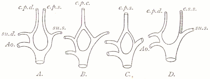
Diagram of some of the variations of the Carotid
Arteries
A.....Normal conditions,
two separate deep carotids;
B.....The two deep
carotids fused into one, e.g. Ardea ;
C.....The same as
B, but the root of the left carotid is reduced, e.g. Phoenicopterus ;
D.....The left deep
carotid is lost, but supplanted by a superficial vessel, e.g. certain Psittaci.
Ao.....Aorta
su.d.....A. subclavia
dextra
su.s.....A. subclavia
sinistra
c.p.d.....A. carotis
profunda dextra
c.p.s.....A. carotis
profunda sinistra
c.p.c.....A. carotis
profunda conjuncta
c.s.s.....A. carotis
superficialis sinistra |
|
3. There is one carotis conjuncta, but
the right root, i.e. the basal portion of the original right carotis,
has been obliterated. The artery is a so-called "carotis primaria sinistra."
Such "Aves laevocarotidinae"
(as called by Garrod) are very frequent,
e.g.
Rhea and Apteryx
among the Ratitae, Podicipes, several Steganopodes, Alca, Otis, Turnix,
Megapodiidae, some Old-World Psittaci, Merops, Buceros, Upupa, Trogonidae,
Cypselidae, Colius, all the Pici and Passeres.
4. One carotis conjuncta, but the right root alone
is present, the left being obliterated. "This carotis primaria dextra"
is likewise deeply lodged, as in the 2nd and 3rd cases, and has hitherto
been observed only in Eupodotis.
In the following three cases, one or two collateral
and superficially-placed arteries take the place of one or both deep carotids.
5. A carotis primaria s. profunda dextra
coexists with a carotis superficialis s. collateralis sinistra. All the
American and a few Old.WorId Parrots are such "Aves bicarotidinae abnormales"
(Garrod).
6. Two superficial carotids, a right and left,
are present, the deep or primary vessels being entirely obliterated. This
was observed by OttIey (P.Z.S. 1879, p. 461), as an individual variation
of Bucorvus abyssinicus.
7. The only carotis is a carotis superficialis
sinistra, all the other vessels being lost, observed by Forbes in Orthonyx
spinicauda (not in O. ochrocephala), this being the only exceptional case
of all the Passeres examined at his time.
It is clear that the 2nd case is directly referable
to the 1st, that the 3rd and 4th are each independently developed from
the 2nd, and that the 5th, 6th, and 7th cases are recent and very qualified
modifications. The undoubtedly independent acquisition of these carotid
characters renders them valueless for taxonomic purposes, except within
smaller and well-defined groups, e.g.
the Parrots (see also VASCULAR
SYSTEM).
|
|


