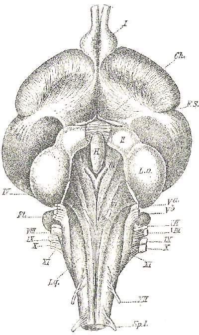
Diagram of ventral view of the
brain structure of a Goose
I-XII.....The
twelve pairs of cranial nerves
Ch.....Chiasma of
the optic nerves cut across
Fl.....Flocculus
H.....Hypophysis
L.o.....Optic lobe
Lq.....Laqueus
F.S.....Sylvian fissure
Sp.I.....First spinal
nerve
|
I. The Olfactory Nerve (Nervus olfactorius)
forms the anterior and ventral continuation of the hemisphere of its side,
but arises in reality from ganglionic cells in the thalamencephalon and
the midbrain. It leaves the cranial cavity through a canal in the dorsal
and median part of the orbit and ends in the ganglionic cells of the olfactory
membrane of the nose.
II. The Optic Nerve (Nervus opticus) arises
from the ganglionic cells of the mantle of the optic lobes. Immediately
in front of the hypophysis is the optic chiasma, produced by the complete
crossing of the fibres, which compose the two optic nerves, those from
the right optic lobe passing over the left, and those from the left lobe
to the right side. From the chiasma start the right and left optic nerves,
each leaving the cranium by the large optic foramen between the orbitosphenoid
and alisphenoid, entering the orbit near the posterior and ventral corner
of the orbital septum and ultimately forming the retina of the eye.
III. The Oculomotor Nerve (Nervus oculomotorius)
arises close behind the hypophysis, near the medio-ventral line, from
the midbrain, enters the orbit behind or together with the optic nerve
(II), and supplies most of the external muscles of the eye, namely the
m. rectus superior, inferior, internus, and obliquus inferior. A ciliary,
partly sympathetic, branch supplies the eyeball and the internal muscles
(see EYE).
IV. The Trochlear Nerve (Nervus trochlearis
or patheticus) is the only one which leaves the brain on its dorsal surface,
namely as a thin thread winding its way from the midbrain upwards between
the cerebellum and the optic lobes, and entering the orbit through a fine
opening close to the optic nerve (II) in order to supply the m. obliquus
superior of the eyeball.
V. The Trigeminal Nerve (Nervus trigeminus)
is next to the optic the thickest nerve, and of a complex nature, being
motory and sensory. It arises from the sides of the midbrain and hindbrain,
forms the large Gasserian ganglion in the wall of the cranium, and leaves
the latter in the form of three branches.
-
The first or ophthalmic branch comes directly out
of the ganglion through a foramen behind the optic (II), runs along the
dorsal corner of the orbital septum, and leaves the orbit at its inner
anterior corner in order to supply the palate, the bill, forehead, and
the lacrymal gland. It is chiefly sensory, and consequently strongest in
birds with tactile bills, like Ducks and Snipes.
-
The second or upper maxillary branch runs along the
ventral edge of the orbital septum, and besides the palatine and maxillary
regions supplies the eyelids and Harder's gland.
-
The third or inferior maxillary branch is the strongest
of the three; it leaves the cranium together with the second through a
foramen between the basi-alisphenoid and petrosal bones and innervates
all the masticatory muscles, the parotid gland, and the whole of the under
jaw.
VI. The Abducens Nerve (Nervus abducens)
is a very thin nerve arising from the hindbrain near the medio-ventral
line, entering the orbit through a special foramen latero-ventrally from
the optic foramen, and supplying the lateral rectus muscle and the two
muscles of the nictitating membrane. It is entirely motory.
VII. The Facial Nerve (Nervus facialis)
arises from the side of the hind brain, possesses a ganglion (known as
the ganglion geniculatum), passes through the petrosal bone into the Fallopian
canal, and sends the sympathetic sphenopalatine branch to the second branch
of the trigeminal nerve (V). The facial nerve leaves the tympanic
cavity behind the quadrate bone, supplies the digastric muscle or depressor
of the mandible, the little stapedius muscle of the ear-bones, the mylohyoid
and stylohyoid muscles of the tongne, and further on connects itself with
branches from the first four cervical nerves and occasionally with branches
from the glossopharyngeal nerve (IX), ultimately supplying the skin on
the front of the neck. There are no branches, as in Mammals, to supply
the face, nor is there in Birds a chorda tympani, i.e. a branch of the
facial nerve joining the mandibular branch of the trigeminal nerve (V).
VIII. The Vestibulocochlear Nerve (Nervus
acusticus) arises dorsally from the facial nerve (VII), of which
it is the sensory portion. It is very short and thick, possesses a little
ganglion, and spreads out in the cochlea of the EAR as the nerve of hearing.
|